Anatomy, Physiology
 https://www.youtube.com/c/Anatomyzone/playlists
Anatomy
https://www.youtube.com/c/Anatomyzone/playlists
Anatomy
Summary of the physical structures of the body, focused on the limits of current research. The goal is to outline a system of development, enabling the activation and use of lesser-understood or presently undiscovered systems.
Body - Foundation
Investigate (inquire, look at, 'ask') physical structure, with an eye towards seeing connctions and establishing valid correspendences.
Hospital Departments
Medical School
Neurophysiology
Brain Structure, Function
Brain Mapping, Neurology
Pineal - Light, Melatonin, Piezo-electric crystals, 3rd Eye, Eye of Horus, mutidimensional, transciever, transducer, radio waves, EMF, calcification, restoration, developmental exercises.
Spinal cord, spinal coumn, 'Jacob's Ladder'
The OLDER a word is, not only did it have more time to develop and gather power of meaning and usage, but also to grow more varied associations of meaning.
We can make analogies between physical structures, their function, and an extended or REVEALED structure that you guess or postulate to be there, guided by morphology in the same way we make assertions about fruit medicinal properties from the nature of their skin.
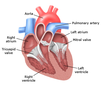
The Heart
The heart has four valves, namely the mitral, aortic, tricuspid, and pulmonary, that regulate the flow of blood through the heart's four chambers. Each valve consists of a flap, or leaflet, that regulates the blood flow to adjacent chambers, then snaps shut to prevent blood from flowing backwards. As in an automobile engine, valves can experience leakage, a situation in which valves do not close completely, allowing blood to flow in reverse. A second valve disorder is stenosis, in which the malfunctioning valve limits the volume of blood flow.
Both conditions can significantly reduce the heart's ability to pump blood. In many cases, heart disease progresses slowly, as the heart compensates for irregularities in blood flow, so symptoms may not seem severe. One may appear symptom-free, yet have serious heart valve disease, requiring immediate hospitalization. In general, irregular valve activity creates abnormal heart sounds, such as murmurs and clicks, that can be heard with a stethoscope. Finally, an echocardiogram may be called for in order to confirm the diagnosis. Further diagnostics can be performed, such as CT-angiography and cardiac MRI.
Anatomy of the Lungs
The lungs have miles of tiny passages, easily clogged by pollutants such as smoke, and other microscopic irritants. Asthma is a chronic lung disease that narrows the airways. In the US, more than 25 million people are known to have asthma, and new research indicates that a chemical compound found in many air fresheners, bathroom cleaners, and deodorizing products, may be harmful to the lungs.
Air first enters your body through your nose or mouth, which wets and warms the air. Conversely, cold, dry air can irritate your lungs. The air then travels through your voice box and down your windpipe, which splits into two bronchial tubes entering the lungs. A thin flap of tissue called the epiglottis covers your windpipe when you swallow, preventing food and drink from entering the air passage.
Except for the mouth and some parts of the nose, all of the airways are covered by cilia, which contain a sticky, mucus coating. The cilia trap germs and other foreign particles that enter your airways when you breathe in. Fine hairs then sweep the particles up to the nose or mouth. From there, they're swallowed, coughed, or sneezed out of the body.
Your lungs and associated blood vessels deliver oxygen to your body and remove carbon dioxide. Interestingly, the left lung is slightly smaller than the right lung, allowing additional room for your heart. Within the lungs, individual bronchi branch into thousands of thinner tubes called bronchioles. These tubes end in bunches of tiny round air sacs, the alveoli. Each air sac is covered by a mesh of tiny capillaries. The pulmonary artery delivers blood rich in carbon dioxide (lacking in oxygen) to the capillaries that surround the air sacs. Inside, carbon dioxide migrates from the blood back into the air. At the same time, oxygen is absorbed. The oxygen-rich blood then travels to the heart through the pulmonary vein, completing respiration.
The Digestive System
The digestive system is made up of the alimentary canal (also called the digestive tract) and other organs, such as the liver and pancreas. The alimentary canal is the long tube of organs including the esophagus, stomach, and intestine. An adult's digestive tract is about 30 feet (about 9 meters) long.
As the teeth tear and chop the food, spit moistens it for easy swallowing. A digestive enzyme in saliva called amylase (pronounced: AH-meh-lace) starts
to break down some of the carbohydrates (starches and sugars) in the food even before it leaves the mouth.
Swallowing, done by muscle movements in the tongue and mouth, moves the food into the throat, or pharynx (pronounced: FAIR-inks). The pharynx is a passageway for food and air. A soft flap of tissue called the epiglottis closes over the windpipe when we swallow to prevent choking.
From the throat, food travels down a muscular tube in the chest called the esophagus. Waves of muscle contractions called peristalsis force food down through the esophagus to the stomach. A person normally isn't aware of the movements of the esophagus, stomach, and intestine that take place as food passes through the digestive tract.
At the end of the esophagus, a muscular ring allows food to enter the stomach and then squeezes shut to keep food or fluid from flowing back up into the esophagus. The stomach muscles churn and mix the food with digestive juices that have acids and enzymes, breaking it into much smaller, digestible pieces. An acidic environment is needed for the digestion that takes place in the stomach.
By the time food is ready to leave the stomach, it has been processed into a thick liquid called chyme. A walnut-sized muscular valve at the outlet of the stomach called the pylorus keeps chyme in the stomach until it reaches the right consistency to pass into the small intestine. Chyme is then squirted down into the small intestine, where digestion of food continues so the body can absorb the nutrients into the bloodstream.
The Small Intestine
The inner wall of the small intestine is covered with millions of microscopic, finger-like projections called villi (pronounced: VIH-lie). The villi are the vehicles through which nutrients can be absorbed into the blood. The blood then brings these nutrients to the rest of the body. The small intestine is composed of three parts:
- The duodenum, the C-shaped first part
- The jejunum, the coiled midsection
- The ileum, the final section that leads into the large intestine
The liver (under the ribcage in the right upper part of the abdomen), the gallbladder (hidden just below the liver), and the pancreas (beneath the stomach) are not part of the alimentary canal, but these organs are essential to digestion.
The liver makes bile, which helps the body absorb fat. Bile is stored in the gallbladder until it is needed. The pancreas makes enzymes that help digest proteins, fats, and carbs. It also makes a substance that neutralizes stomach acid. These enzymes and bile travel through special pathways (called ducts) into the small intestine, where they help to break down food. The liver also helps process nutrients in the bloodstream.
From the small intestine, undigested food (and some water) travels to the large intestine through a muscular ring or valve that prevents food from returning to the small intestine.
ANATOMY OF THE EYE
The eye sits in a protective bony socket called the orbit. Six extraocular muscles in the orbit are attached to the eye. These muscles move the eye up and down, side to side, and rotate the eye. The extraocular muscles are attached to the white part of the eye called the sclera. This is a strong layer of tissue that covers nearly the entire surface of the eyeball.
The surface of the eye and the inner surface of the eyelids are covered with a clear membrane called the conjunctiva. Tears lubricate the eye and are made up of three layers. These three layers together are called the tear film. The mucous layer is made by the conjunctiva. The watery part of the tears is made by the lacrimal gland. The eye’s lacrimal gland sits under the outside edge of the eyebrow (away from the nose) in the orbit. The meibomian gland makes the oil that becomes another part of the tear film. Tears drain from the eye through the tear duct.
Light is focused into the eye through the clear, dome-shaped front portion of the eye called the cornea. Behind the cornea is a fluid-filled space called the anterior chamber. The fluid is called aqueous humor. The eye is always producing aqueous humor. To maintain a constant eye pressure, aqueous humor also drains from the eye in an area called the drainage angle. Behind the anterior chamber is the eye’s iris (the colored part of the eye) and the dark hole in the middle called the pupil. Muscles in the iris dilate (widen) or constrict (narrow) the pupil to control the amount of light reaching the back of the eye.
Directly behind the pupil sits the lens. The lens focuses light toward the back of the eye. The lens changes shape to help the eye focus on objects up close. Small fibers called zonules are attached to the capsule holding the lens, suspending it from the eye wall. The lens is surrounded by the lens capsule, which is left in place when the lens is removed during cataract surgery. Some types of replacement intraocular lenses go inside the capsule, where the natural lens was. By helping to focus light as it enters the eye, the cornea and the lens both play important roles in giving us clear vision. In fact, 70% of the eye's focusing power comes from the cornea and 30% from the lens.
The vitreous cavity lies between the lens and the back of the eye. A jellylike substance called vitreous humor fills the cavity. Light that is focused into the eye by the cornea and lens passes through the vitreous onto the retina, the light-sensitive tissue lining the back of the eye. A tiny but very specialized area of the retina called the macula is responsible for giving us our detailed, central vision. The other part of the retina, the peripheral retina, provides us with our peripheral (side) vision.
The retina has special cells called photoreceptors. These cells change light into energy that is transmitted to the brain. There are two types of photoreceptors: rods and cones. Rods perceive black and white, and enable night vision. Cones perceive color, and provide central (detail) vision. The retina sends light as electrical impulses through the optic nerve to the brain. The optic nerve is made up of millions of nerve fibers that transmit these impulses to the visual cortex, the part of the brain responsible for our sight.
The Ear
The ear depends on coordinated events that transform sound waves into electrical impulses. The auditory nerve transmits these signals to the brain. Initially, sound waves enter the outer ear and traverse the outer ear canal, leading to the eardrum. The eardrum vibrates from the incoming sound waves and sends vibrations to three tiny bones in the middle ear.
These bones couple the sound waves from the air to fluid vibrations in the cochlea of the inner ear. Hair-like sensory cells perched on top of the basilar membrane ride the ripple of fluid thus created. As the hair cells move up and down, microscopic stereocilia sitting on top of the hair cells bump against an overlying structure and bend, which causes pore-like channels at the tips of the stereocilia to open. When that happens, chemicals rush into the cell, sparking an electrical signal. The auditory nerve then carries this signal to the brain, which translates it into a sound that we can recognize and understand.
When exposed to loud noises over an extended period, hearing losses may occur. Over time, sounds become distorted, and it may be difficult to understand other people when they talk. Sometimes exposure to continuous noise causes a temporary hearing loss, but there also may be residual long-term damage. Loud noise exposure also may be responsible for tinnitus, which is perceived as a ringing in the ears or cranium.
Biological scientists used 3-D printing of cartilage cells and nano-sized materials to create functional ears that receive radio signals. The experiments demonstrated that it may be possible to create bionic tissues and organs. Scientists used 3-D printing to merge living tissue with an antenna that is able to receive radio signals. In tissue engineering, cells and other biological materials are used to augment or replace deteriorating muscle matter, bone and cartilage. Currently, it’s difficult to create 3-D structures for use in the body, especially organs with complex geometry such as the ear.
*****BRAIN
- Neurons
- Neurotransmitters
Spinal Chord
Cardiac Plexus
Pulmonary Plexes
Solar Plexus
Pelvic Plexus
Nervous System
Nerves
** SKIN, hand, touch - organ of perception
** Bones - Feel it in my Bones
(Bones are roots, Bone Marrow, Stem Cells)
** Digestion
Second Brain - organ of perception
Intuition - Gut Feeling (RIGHT BRAIN)
Gut-Mind connection
** Ear - audio - hear - understand
** Eye - Optical - see - to undrstand
Hormonal System
Pineal - Third Eye - organ of perception
Hypothalumus - Control Organ, Regulation, Homeostasis
Adrenal Glands
Pituitary Gland
Skin
Integumentary System
** SKIN - organ of perception
Touch, In Touch, Touchy (sensitive), Picky (particular), Pick-up (to date)
eat food through the skin, acupuncture points
Skin
Lymphatic System
Immune System
Gut
Lymphocytes
Muscles
Fiber Contraction
HEART, BLOOD, ELECTRICITY
1. Heart - Cardiology, Medical Experimentation
2. Look at the Heart symbolically, representationally, making logical associations, using language keys, tips from morphology, function, myth, analogy, Metaphor, stories, other researchers, ancient texts, ancient structures (figure it out)
3. 'Seeing' the 'heart' spiritually (Third Eye), asking higher sources
CARDIO-PULMONARY SYSTEM
CIRCULATORY SYSTEM
Heart - Cardiac - the heart of the matter, Love
Frank Chester - regular 7-sided polygon, sitting in a box, at 36 degrees, like a heart - spin in water, fluid vortex, plasma, structure water (blood)
Francisco Torrent-Guasp - helical band of msucle
http://www.torrent-guasp.com/pages/Research.htm
Biomaterials
Bio-energetics, EMF, plasma, sound pulse
Anatomical Structure
Cardiac Functions
-
-
-
Water
Viktor Schauburger
Emoto
Blood - Circulatory System
Polarity, charge, ions, salts
Electrostatics
Circulation
Quantum EMF generator
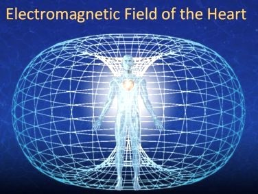
HEART CHAKRA
Pulsating Magnetic Field
Blood, Oxygen (breathing)
Cardiac Plexus
Field Strength
SQUID magnetometers
heart's magnetic field (magnetocardiogram)
Respiration
Food
LUNGS, AIR, OXYGEN, PRANA
Lungs - Pulmonary
Control Systems
Regulation
Neural
Hormonal - Pineal, Pituitary, Hypothalmus, Adrenal
Meridianal - Acupuncture
Cleansing, Purification, Detox
Rejuvenation
Lymph
Kidneys
Liver
Lungs
Digestion, Absorbtion
skin - receiver, reception
teeth - resonators, key points in acupuncture
Stomach - ruminate in the gut, turn it over, I'm still digesting that information, I am taking it in
intestines - absorbtion, antenna
VISCERAL BRAIN - Internal Awareness ad Understanding, Ancient Intuition
Digestion, Gut
- Second Brain, Fasting
Reproduction
root - reproduction
If you can make it from scratch, you can repair it, and can keep it in working order.
Bone
SOUND, Vibration
Research tests with various classical music with plants, which grew fastest. look at the winners, what notes and keys were the pieces written in?
We may use the bones to resonate, vibrate, respond to frequency or sound input, in order to broadcast a signal through the bones into the body.
Broadcast sound into the body. Tartaria
Bone - center of the body, in a way.
Bone marrow - center of the bone
Composed of STEM CELLS
Red Blood Cells
Lymphocytes
In the structure of a bone, the primary ions present as charged particles are calcium (Ca2+) and phosphate (PO4^3-) ions which together form the mineral hydroxyapatite, the main component giving bone its rigidity and strength; other ions like magnesium (Mg2+) and sodium (Na+) may also be present in smaller quantities contributing to the overall bone composition.
DNA
DNA, the ancient cabalistic "Tree Of Life" portrayed in the Biblical Torah, is now coming to be viewed as a live vibrating structure, rather than a fixed tape recording.
Many modern scientists, regard DNA as a shimmering, waveform configuration, able to be modified by light, radiation, magnetic fields or sonic pulses. The legacy of Thoth/Enoch suggests this "language of Light", the harmonic science of the ancients, could actually affect DNA.